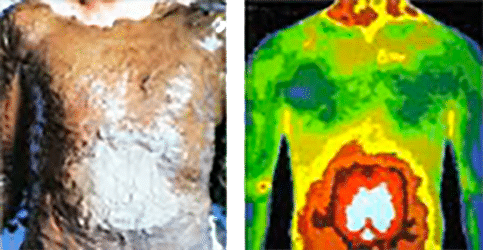
The roots of Thermography, or heat differentiation, are ancient, dating back to the time of the pyramids. A papyrus from 1700 BC documents the association of temperature with disease.
By 400 BC, physicians commonly employed a primitive form of Thermography: they applied a thin coat of mud to a patient’s body, observed the patterns made by the different rates of mud drying, and attributed those patterns to hot and cold temperatures on the surface of the body. Hippocrates summed it up: “In whatever part of the body excess of heat or cold is felt, the disease is there to be discovered.”
Fast forward 2,000 years and thermography is gaining more and more acceptance in the medical world.
Here, at the Family Naturopathic Clinic, we consider thermography as a very important and unique “Health Discovery Technology” that provides thousands of patients with peace of mind when they may be questioning new or unusually concerning health symptoms.
Skin blood flow is under the control of the sympathetic nervous system. In normal people there is a symmetrical dermal pattern which is consistent and reproducible for any individual. This is recorded in precise detail with a temperature sensitivity of 0.1 Celsius with our scanner.
In addition, thermography can assist in the discovery of the state of health in general. You may have heard of thermography as a safe method of breast health screening…but it can be that and so much more!
Role in Breast Cancer screening
Thermography is a painless, non-invasive, state of the art clinical test without any exposure to radiation and is used as part of an early detection program which gives women of all ages the opportunity to increase their chances of detecting breast disease at an early stage. It is particularly useful for women under 50 where mammography is less effective.
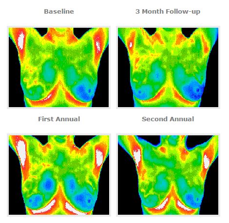
Thermography’s role in breast cancer and other breast disorders is to help in early detection and monitoring of abnormal physiology and the establishment of risk factors for the development or existence of cancer. When used with other procedures the best possible evaluation of breast health is made.
This test is designed to improve chances for detecting fast-growing, active tumors in the intervals between mammographic screenings or when mammography is not indicated by screening guidelines for women under 50 years of age.
All patients thermograms (breast images) are kept on record and form a baseline for all future routine evaluations.

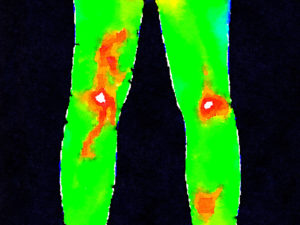
Abnormal presentation of the legs
To sum up, the clinical uses for Infrared Thermal Imaging include:
- To define the extent of a lesion of which a diagnosis has previously been made;
- To localize an abnormal area not previously identified, so further diagnostic tests can be performed;
- To detect early lesions before they are clinically evident;
- To monitor the healing process before the patient is returned to work or training.
Over the past two decades, it has been determined through extensive clinical studies that inflammation is not just a result of sickness, but the actual CAUSE of serious, life threatening conditions like heart disease, cancer and diabetes. Inflammation, most often initially caused by the bio-chemical results of stress, creates an environment that allows these kinds of diseases to establish and flourish. This condition is silent but not invisible… if you can just put yourself in front of an infrared camera and an experienced thermographer. This is why you should use thermography… knowledge offers the power to take control of your future health and the length and quality of your life.
